MRI vs X-Ray What’s the Difference and When Is Each Used?
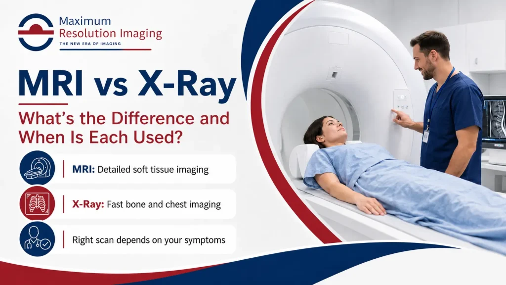
MRI and X-Ray are two common imaging tests used to help doctors see what is happening inside the body. Both can provide important information, but they work in very different ways and are often used for different medical reasons.
An X-Ray is usually quick and commonly used to evaluate bones, chest concerns, certain injuries, and some internal conditions. An MRI provides more detailed images of soft tissues, organs, muscles, ligaments, nerves, and joints. The right test depends on your symptoms, the body area being examined, and your provider’s recommendation.
At Maximum Resolution Imaging Center Texas, patients in Richmond, Houston, and nearby areas can access advanced diagnostic imaging services, including MRI scans and X-Rays, in a patient-focused setting.
What Is an MRI?
MRI stands for Magnetic Resonance Imaging. It uses a strong magnetic field and radio waves to create detailed images of the body’s internal structures. MRI does not use ionizing radiation.
MRI is especially helpful when doctors need a closer look at soft tissues. This can include the brain, spine, joints, muscles, ligaments, tendons, organs, and other internal structures. It is often used when symptoms or injuries are too detailed to evaluate clearly with a basic X-Ray.
MRI may be recommended for:
What Is an X-Ray?
An X-Ray is a fast imaging test that uses a small amount of ionizing radiation to create pictures of structures inside the body. X-Rays are commonly used because they are quick, simple, and helpful for many common concerns.
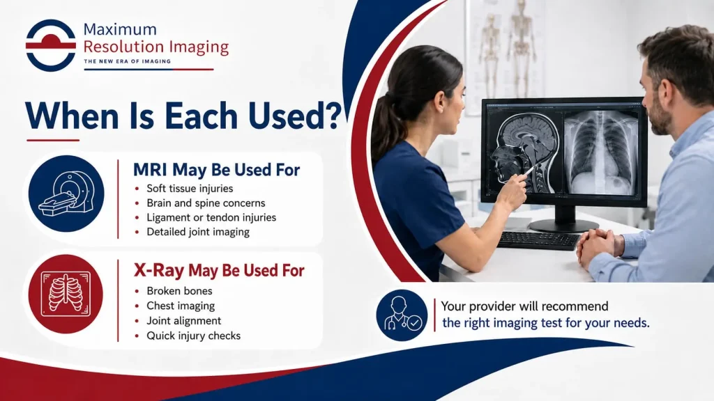
X-Rays are especially useful for evaluating bones and certain chest or abdominal concerns. They may help detect fractures, dislocations, arthritis changes, lung concerns, infections, foreign objects, or other visible abnormalities.
X-Ray imaging may be recommended for:
MRI vs X-Ray Quick Comparison
MRI and X-Ray are both useful imaging tests, but they are used for different reasons. MRI uses magnets and radio waves to create detailed images of soft tissues, joints, muscles, ligaments, nerves, the brain, spine, and organs. It is often recommended when your provider needs a deeper look at areas that may not show clearly on an X-Ray.
X-Ray is usually faster and is commonly used for bones, fractures, chest imaging, joint alignment, and quick injury checks. It uses a small amount of radiation and can help providers quickly identify visible bone or chest-related concerns.
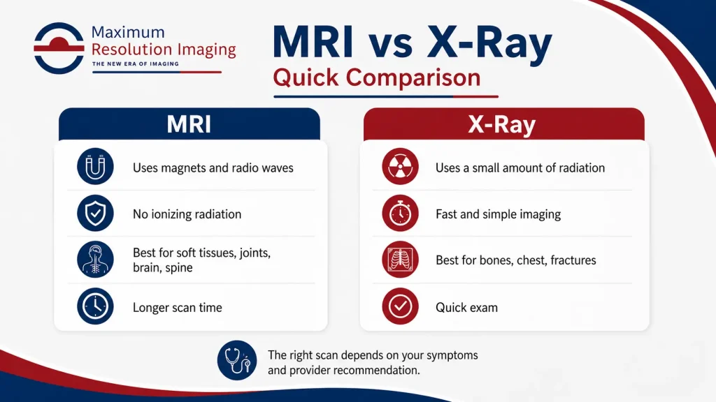
The right choice depends on your symptoms, the body area being checked, and your provider’s recommendation. In some cases, an X-Ray may be used first, and an MRI may be recommended later if more detailed soft tissue imaging is needed.
|
Feature |
MRI |
X-Ray |
|
Imaging Method |
Uses magnets and radio waves |
Uses a small amount of radiation |
|
Best For |
Soft tissues, brain, spine, joints, organs, ligaments, tendons |
Bones, chest, joints, fractures, certain abdominal concerns |
|
Radiation |
No ionizing radiation |
Uses ionizing radiation |
|
Scan Time |
Usually longer |
Usually very quick |
|
Image Detail |
More detailed for soft tissues |
Clear for bones and dense structures |
|
Patient Experience |
Requires lying still inside or near MRI scanner |
Usually fast and simple positioning |
|
Common Use |
Complex pain, soft tissue injury, neurological or joint concerns |
Fractures, chest X-Ray, injury checks, routine diagnostic imaging |
Both tests are valuable. One is not always better than the other. The best imaging test depends on what your provider needs to evaluate.
When Is an MRI Used?
An MRI is often used when your provider needs detailed images of soft tissues or complex internal structures. For example, if you have ongoing back pain, nerve symptoms, a sports injury, unexplained joint pain, or a possible ligament or tendon injury, an MRI may provide more helpful detail than an X-Ray.
MRI is also commonly used for brain, spine, musculoskeletal, abdominal, pelvic, breast, cardiac, and full-body imaging needs. Because MRI can show soft tissue detail clearly, it may help identify injuries or abnormalities that may not appear well on an X-Ray.
Your provider may recommend an MRI if symptoms continue even after an X-Ray looks normal, or when more detail is needed to guide treatment.
When Is an X-Ray Used?
An X-Ray is often the first imaging test used for injuries, bone pain, chest symptoms, or certain quick diagnostic checks. If a provider suspects a broken bone, dislocated joint, chest infection, or spinal alignment issue, an X-Ray may be recommended first.
X-Rays are usually fast and convenient. They can help doctors quickly review bones, joints, lungs, and certain internal structures. For many injury-related concerns, an X-Ray can give useful information without the longer scan time of an MRI.
An X-Ray may also be used before an MRI to rule out a fracture or check for visible bone changes.
Which Scan Shows More Detail?
MRI usually provides more detail for soft tissues, such as muscles, ligaments, tendons, nerves, the brain, spine, and organs. This makes MRI helpful for complex injuries, neurological concerns, and soft tissue evaluation.
X-Ray provides strong detail for bones and dense structures. It is useful for detecting fractures, joint changes, alignment issues, and some chest or abdominal findings.
So, if the concern is a suspected broken bone, an X-Ray may be enough. If the concern involves soft tissue, nerves, discs, ligaments, or deeper internal structures, an MRI may be the better option.
MRI vs X-Ray for Common Conditions
|
Health Concern |
Imaging Test Often Considered |
|
Suspected broken bone |
X-Ray |
|
Suspected broken bone |
X-Ray |
|
Chest or lung concern |
X-Ray |
|
Joint pain after injury |
X-Ray first, MRI if soft tissue injury is suspected |
|
Ligament or tendon injury |
MRI |
|
Herniated disc or nerve compression |
MRI |
|
Brain or neurological symptoms |
MRI |
|
Abdominal or pelvic soft tissue concern |
MRI |
|
Spine alignment issue |
X-Ray may be used first |
|
Ongoing pain with unclear cause |
Provider may choose MRI, X-Ray, or both |
Your provider will decide which test is most appropriate based on your symptoms, exam findings, and medical history.
Is MRI Safer Than X-Ray?
MRI does not use ionizing radiation, which is one reason it may be preferred for some patients and certain types of imaging. However, MRI uses a strong magnet, so safety screening is very important. Patients with pacemakers, implants, metal fragments, or certain medical devices may need special review before having an MRI.
X-Ray uses a small amount of radiation. For many patients, the benefit of getting the right diagnosis is greater than the small radiation exposure. However, you should always tell your imaging team if you are pregnant or may be pregnant before an X-Ray.
Both tests are considered useful when ordered appropriately. Safety depends on choosing the right test for the right reason.
How to Prepare for an MRI or X-Ray
Preparation depends on the scan being performed. For an MRI, you may be asked to remove jewelry, watches, hearing aids, phones, credit cards, hair pins, and clothing with metal. You should also tell the imaging team about implants, devices, surgeries, or any metal in your body. Before your visit, review the preparation for MRI scan guidelines so you know what to remove, what to bring, and how to prepare safely.
For an X-Ray, preparation is usually simple. You may need to remove jewelry or metal objects from the area being scanned. You may also be asked to change into a gown depending on the body area.
Following the instructions from your imaging center can help improve image quality and make your visit smoother.
Can You Need Both MRI and X-Ray?
Yes, some patients may need both. For example, an X-Ray may be used first to check for a fracture or bone alignment issue. If the X-Ray does not explain the symptoms, or if soft tissue damage is suspected, an MRI may be recommended next.
The two tests can work together. X-Ray gives fast information about bones and certain structures, while MRI gives more detail about soft tissues and deeper internal concerns. In some cases, your provider may also recommend a CT scan if faster cross-sectional imaging is needed for a specific medical concern.
MRI and X-Ray Services at Maximum Resolution Imaging Center Texas
Maximum Resolution Imaging Center Texas provides diagnostic imaging services for patients in Richmond, Houston, and nearby communities. Our team helps patients complete imaging in a comfortable outpatient setting with a focus on clear results, patient safety, and a smooth experience.
Whether your provider has ordered an MRI scan or an X-Ray, our imaging team can explain what to expect, help you prepare, and guide you through the process.
Final Thoughts
MRI and X-Ray are both important diagnostic imaging tools, but they are used for different reasons. X-Ray is often fast and helpful for bones, fractures, chest concerns, and many routine imaging needs. MRI is often better for soft tissues, joints, nerves, organs, and more detailed diagnostic questions.
The right choice depends on your symptoms, the body area being examined, and your provider’s recommendation. If you need imaging in Richmond or the Houston area, Maximum Resolution Imaging Center Texas can help you move forward with confidence.
Schedule Your Imaging Appointment
If your provider has recommended an MRI or X-Ray, Maximum Resolution Imaging Center Texas is here to help. Our team provides advanced imaging services with patient comfort and accurate results in mind.